Menu
- Incheon Sejong Hospital
Call Us
- +82-32-240-8000
- +82-1599-6677
- for Questions and Appointments
- Greetings of Hospital Director
- Department & Staff
- Specialized Center
- Specialty Center
- Health Improvement Center
- Endoscopy Center
- Internal medicine Center
- Surgery Center
- General Health Check-Up Center
- Emergency Medical Center
- Rehabilitation Center
- Spine-Joint Center
- Physiological Function Test Center
- Minimal Invasive Operation Center
- Dementia Special Center
- Infection Center
- Center for Bloodless Medicine and Surgery
- Breast and Thyroid Cancer Center
- Center for Precision Medicine
- Heart Transplantation Center
- Women’s Center
- Hospital Information
- Directions for Visitors
- Health Screening Program
- Get your Health Examination!

- Bucheon Sejong Hospital
Call Us
- +82-32-240-8000
- +82-1599-6677
- for Questions and Appointments
- Greetings of Hospital Director
- Department & Staff
- Cardio-Cerebrovascular Center
- Specialty Center
- Hospital Information
- Directions for Visitors
- Health Screening Program
- Get your Health Examination!
- Appointment Service
Call Us
- +82-32-240-8000
- +82-1599-6677
- for Questions and Appointments
- Estimate Treatment Expense
- Estimate Treatment Expense

- Customer Center
Call Us
- +82-32-240-8000
- +82-1599-6677
- for Questions and Appointments
- Estimate Treatment Expense
- Estimate Treatment Expense
- About Foundation
Call Us
- +82-32-240-8000
- +82-1599-6677
- for Questions and Appointments
- Overview
- Chairman's Message
- Mission&Vision
- Medical Service Works
- Promotion Video
- Introduction Brochure
- Incheon Sejong at a glance
- Your very best Total Medical Service valuing clients and patients

- home
- Incheon Sejong Hospital
- Specialty Center
-
- Health Improvement Center
- Endoscopy Center
- Internal medicine Center
- Surgery Center
- General Health Check-Up Center
- Emergency Medical Center
- Rehabilitation Center
- Spine-Joint Center
- Physiological Function Test Center
- Minimal Invasive Operation Center
- Dementia Special Center
- Infection Center
- Center for Bloodless Medicine and Surgery
- Breast and Thyroid Cancer Center
- Center for Precision Medicine
- Heart Transplantation Center
- Women’s Center

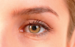
Taking responsibility of the optic health of 3million inhabitants of Incheon
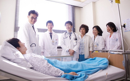
Experience a more professional medical service at Incheon Sejong Hospital which induced the Ministry of Health and Welfare-designated Eye Specialized institute, Optic Hospital system, where annually more than 200,000 patients visit, exactly the way it was. Our Board of Staff who perform treatment consists of five professors of university hospital including someone who has held position as head of the Korean Ophthalmological Society, all specializing in their fields in ophthalmalogy, and we are providing the finest medical service for patients with severe cardio-cerebrovascular diseases with a background of specialized know-hows on patients in need of highly strenuous eye surgery.
Anterior Segment clinic
We treat cataracts, dry eye syndrome, pterygia, conjunctivitis and blepharitis.
Cataract is a condition where one's sight slowly deteriorates as the crystalline lenses become unclear, and it is on constant increase among the elderly due to the lengthening average span of life.
Other than aging, trauma, a longterm usage of steroids and retinitis pigmentosa can be factors of secondary cataract.
The cataract ultrasound surgery, or phacoemulsification of cataract, is an effective way of treatment, with very quick recovery in eyesight, and includes a process of topical anesthesia and retrobulbar anesthesia along with an incision of about 3mm in size, and does not need a suture after the surgery. Our Board of Staff, which include professors of university hospital including a retina-expert who has held position as head of the Korean Ophthalmological Society, will provide with the most accurate and appropriate to textbook finest medical service for patients.


Retinal Clinic
At the Retinal Clinic, we treat diseases such as diabetic retinopathy, which is a major factor of acquired blindness, macular degeneration, and others including retinochorioiditis centrali, retinal vein occlusion, uveiti, muscae volitantes( flying flies) and retinal detachent.
With the center being equipped with the latest equipments, a wider range of screen testing is facilitated, compared to the former which was capable of basic fundus photography, with a wide-range fundus photography, a wide-range vascular angiography, spectralis OCT, contactless electroretinography and visual evoked potential test, effective diagnosis of retinal diseases can be performed.
Treatments, such as intravitreous injection of antibodies, steroid injection, panretinal photocoagulation (PRP) and vitrectomy, are performed by retina experts.
Glaucoma Clinic
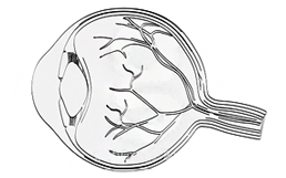
Glaucoma is a disease in which the nerve fibers of the optic nerve, which plays a crucial role in transferring the information from the eye to the brain, get damaged and result to a narrowing of sight.
An alerting aspect of glaucoma is that in many cases it shows no symptom even to its final stages where loss of sight occurs, making it heart to find if not for early diagnosis.
Even for diagnosing, equipments which require precision to minuteness are needed, for a process of visual field testing, optical coherence tomography (OCT) and nerve fiber layer photography, for a definite diagnosis or glaucoma.
Treatment methods such as drug treatment, laser iridotomy and laser trabeculectomy are available, and precise diagnosis and treatment is possible, with the center being equipped with the latest facilities like Humphrey Field Analyzer, spectralis OCT and cirrus OCT, along with a specialist in glaucoma treating regularly.

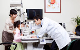
Strabismus·Pediatric Ophthalmology Clinic
For growing children, in order to prevent a decline in sight, it is important for them to adjust a pair of glasses when a certain level of symptom of short-sightedness, long-sightedness and astigmatism are seen.
If vision correction via glasses misses the appropriate timing, problems in sight development might occur and thus result to a state of unredeemable amblyopia.
Therefore adjustment via glasses of growing children must be consulted with an expert.
Strabismus is a condition in which the eyes do not properly align with each other when looking at an object, and this causes esthetic problems.
In cases of children, negligence of strabismus can lead to amblyopia and deterioration in view of three-dimensional objects, double vision, dizziness and headaches.
At Eye Center, specialists in strabismus·pediatric ophthalmology prescribes glasses, occlusive treatment, prismatic spectacles and strabismus surgery take charge of professional treatment.

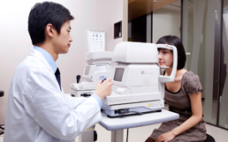
Basic Test Equipments
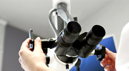
Slit lamp Microscope
Used to differentiate diseases and observe state and structure of anterior segment such as conjunctiva, cornea, the anterior chamber, crystalline lenses, anterior vitreous body and even classify location of lesion to precision. A graphic observation of anterior chamber angle, ocular fundus, posterior vitreous body as well as the retina can be seen when using substitutional lenses.
Cornea Specular Microscope
The equipment used to speculate endotheliocytes in the cornea. Endotheliocytes play an important role in maintaining the clarity of the cornea, and their number is extremely important in diagnosing ophthalmic illnesses, especially for the cornea, which gets easily damaged from different conditions and surgeries, and do not regenerated once damaged.
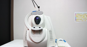
Fundus Camera
The equipment for basic ophthalmic tests such as sight and intraocular pressure testing, which is a test to observe the center of the retina, at the inmost part of the eye, to decide for abnormalities like cataratcs, glaucoma, retina related illnesses, via a fundus photography. At the Eye Center we are equipped with the latest wide-range fundus camera leading a further step into fundus photography by enabling to a wide range of photograph that allows to check an almost complete sight of the retina, thus allowing accurate diagnosis without miss.
Tonometer
The most basic test to diagnosing ophthalmologic diseases, tonometry is most important for diagnosing and treatment of glaucoma. At the center we seek for an accuracy of 100% in measuring intraocular pressure by utilizing 3 methods of tonometry, namely noncontact tonometry, rebound tonometry and applanation tonometry.
Special Test Equipments
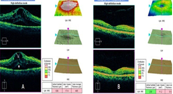
OCT(Optical Coherence Tomography)
With this equipment we can discover minute lesions that might have been missed in the fundus test.
A high-density OCT equipment is needed in order to accurately monitor glaucoma and search for location of lesion in paramacular degeneration.
At the Eye Center, the latest model of both spectralis OCT and cirrus OCT are equipped and so precise diagnosis of glaucoma and retinal illnesses can be made.
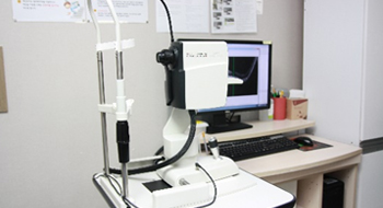
Wide FAG & ICG (Fluorescein Fundus Angiography)

The equipment used to get a grasp of retinal vascular abnormalities, retinal pigment epitheliopathy and choroidal vascular abnormalities, it is used to determine the necessity of light coagulation treatment, to assemble a treatment plan including fluorescent dye for diagnosis of fundus diseases, and also used for evaluating treatment effects.
Humphrey Visual Field (VF)

The equipment used to examine change in the visual field for diagnosis of glaucoma, for locating lesions in the visual pathway and to gather information important for observation of disease progress.
ERG (Measuring response of retina to light, Electroretinography)
Equipment used to roughly decide the function of cornea when fundus examination is impossible due to corneal opacity, cataracts, iridolysis, vitreous opacity and such, Our center induced the ERG, which is usually equipped in hospitals of university-hospital level, in order to precisely diagnosis retinal diseases.
- This is when ther are usually used.
-
- LASIK and LASEK sight correcting surgeries, cornea transplants, estimating prognosis of sight restoration before and after cataract surgery.
- To get a grasp of retinal function when fundus examination is impossible due to corneal opacity, cataracts and vitreous opacity.
- When retinal observations such as retinitis pigmentosa is possible but an examination of retinal function and definite diagnosis, and evaluation of retinal dysfunction is necessary.
- For diagnosis and estimating prognosis of various retinal diseases.
VEP (Response measurement of the cerebral and optic nerve to light, Visual Evoked Potential)
A study to record the response of central nerve system when the optic nerves are stimulated. It is also compulsory equipment needed to judge disorders of optic nerve, as a set of computerized standardization of responses of nervous system to sensory stimuli.
- This is when ther are usually used.
-
- For diagnosis of optic nerve disorders
- For diagnosis of visual pathway disorders
- For diagnosis of corneal opacity, cataracts, vitreous opacity when findings from fundus examination and ophthalmoscopy are insufficient or unclear
- To evaluate visual function of infants and children whose visual acuity cannot be measured
- To evaluate visual function pre-corrective vision surgeries such as LASIK or LASEK
Yag laser
When after a cataract surgery, dust-like clouding occurs on the thin membrane of posterior capsule, left in order to keep the artificial lenses fixed, sight deterioration may occur. Here, we can use YAG laser for a more simple method of devising betterment for sight deterioration, not to mention promoting a speedier recovery of patient's sight.
Pattern laser MC-500V Vixi
Disorders in the retina such as blood vessel wall break, detachment, varix, hemorrhage, edema, effusion, neovascularization and others can directly affect sight. The pattern laser is an equipment that can be used t3o intensify the weak area in the retina, stop hemorrhage in the capillaries using light-coagulation, selective treatment of retina tissue and preventative procedures.
Structure of Eye Center
- Sejoing Hospital Group